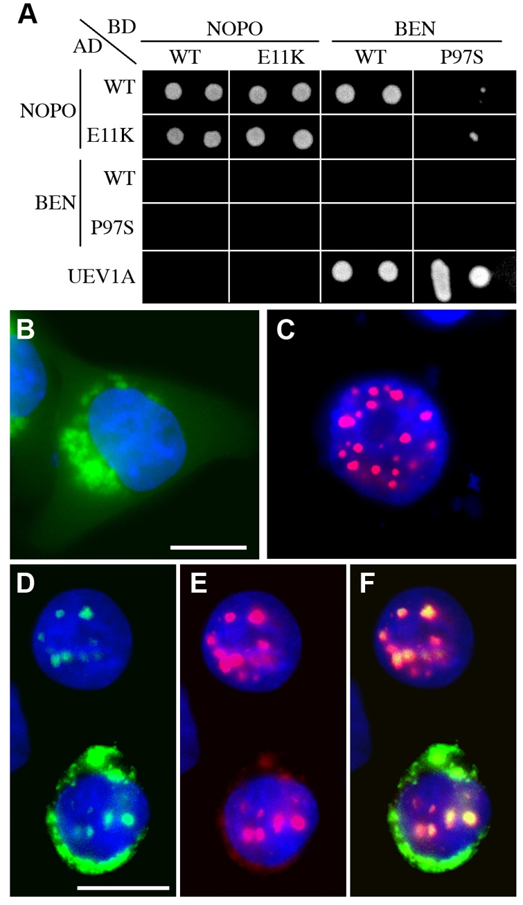
Fig. 6.

NOPO/TRIP, BEN and UEV1A interactions and co-localization. (A) Yeast two-hybrid assay. Yeast cells expressing combinations of NOPO, BEN and UEV1A fused to the Gal4 DNA-binding domain (BD, `bait') or activation domain (AD, `prey') were spotted onto selective media. Growth on SC-Trp-Leu-His media (shown) indicates physical interaction between the fusion proteins. Wild-type and mutant versions of NOPO and BEN (E11K and P97S, respectively) were tested. A representative plate spotted in duplicate is shown; identical results were obtained for three independent Trp+Leu+ colonies per plasmid combination tested. (B-F) Immunofluorescence microscopy of transfected HeLa cells. eGFP-BEN is in green, mCherry-TRIP in red, and DNA in blue. eGFP-BEN (B) and mCherry-TRIP (C) localize distinctly when transfected alone. (D-F) Co-transfection of eGFP-BEN (D) with mCherry-TRIP (E) promotes its localization into nuclear puncta (F, merge). Scale bars: 20 μm.
