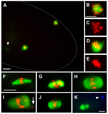
Fig. 7.
ben phenocopies nopo. Representative mitotic spindles in syncytial embryos from wild-type, nopo and ben females. (A-J) Microtubules are in green and DNA in red. (A-E) Single mitotic spindle and polar body in a ben1-derived embryo. (A) Dashed line outlines the embryo; arrowhead indicates detached centrosome out of focal plane. (B-E) Magnified images of the polar body (B,C) and mitotic spindle (D,E) from A. (F-K) Mitotic spindles in embryos from wild-type (F), nopoZ1447 (G), ben1 (H,I) and ben1/Df(1)HA92 (J,K) females. ben-derived embryos exhibit nopo phenotypes, including barrel-shaped, acentrosomal spindles and displaced DNA (I, arrow). (K) Microtubules are in green and centrosomes in blue. A ben1/Df(1)HA92 spindle with a detached centrosome (arrowhead). Scale bars: 20 μm in A; 10 μm in B-K.

