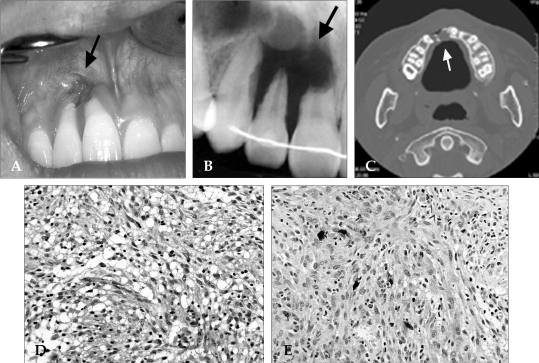
Fig. 3.
A clinical intraoral picture (A) shows a bony depression, gingival recession and root exposure (indicated by arrow) on the right maxillary incisors. A dental radiographic image (B) and maxillary CT (C) reveal an extensive osseous defect and external root resorption of the right maxillary incisors and canine. The elongated tumor cells in the interlacing bundles with surrounding collagen bundles and multinucleated giant cells with abnormal mitoses are seen in H-E staining (D) and they are positive for S-100 protein (E).

