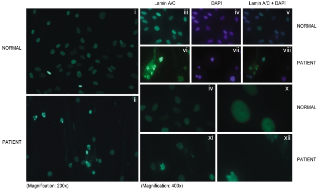
Figure 3.
Indirect immunofluorescence microscopy using lamin A/C antibody reaction in the skin fibroblasts obtained from a control subject (Normal), and the patient. Panels i, ii, iii and vi show immunostaining for lamin A/C; panels iv and vii show nuclear DNA staining; panels v and viii show the merged images from the lamin A/C and DNA staining. Multilobulated nuclei were observed more frequently in the patient. Panels ix, x, xi and xii also show several individual nuclei from the patient, and the nuclei were stained for lamin A/C protein; they displayed various deformities (xi, xii).

