Figure 3.
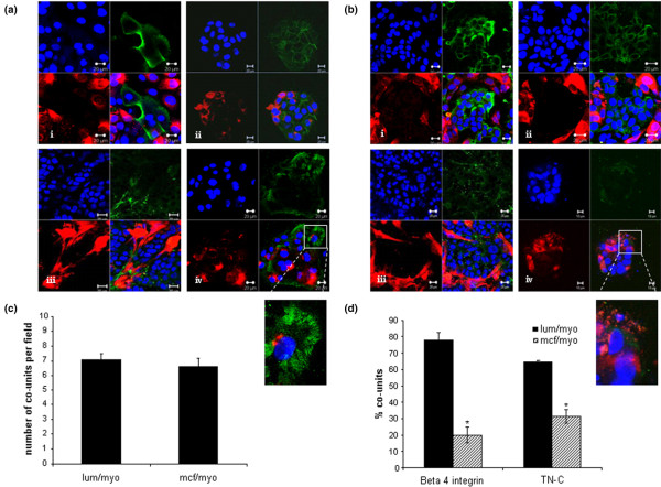
Phenotypic analysis of co-units. (a) Co-units formed by primary myoepithelial cells (red) surrounding the normal luminal epithelial cells exhibited polarity detected by luminal expression of epithelial membrane antigen (EMA) localised to (a)i, green stain: the epithelial population. (a)ii, green stain: E-cadherin was expressed at cell-cell junctions. (a)iii, green stain: Myoepithelial cells laid down the matrix protein tenascin-C (TN-C) and (a)iv, green stain: expressed β4-integrin in hemidesmosome like structures at the myoepithelial-gel interface. (b) In the myoepithelial/MCF-7 co-units, (b)i, green stain: MCF-7 tumour cells expressed EMA and (b)ii, green stain: E-cadherin. A less organised basement membrane was present, (b)iii, green stain: shown by TN-C staining and (b)iv, green stain: marked down-regulation or loss of β4-integrin expression was observed. Quantitation of co-unit formation was carried by counting the number of co-units in 10 microscopic fields per culture to give an average number of co-units/field. A co-unit was defined as aggregated luminal or MCF-7 cells which were at least 70% enclosed by myoepithelial cells. (c) No significant difference in the two types of co-units was observed. To quantitate the presence of β4-integrin and TN-C the percentage of co-units expressing the proteins were counted and expressed as a percentage of total co-units. (d) A significant decrease in the levels of β4 integrin and TN-C were observed in the MCF-7-myoepithelial cell cultures compared with the luminal-myoepithelial cell cultures. (p = 0.001).

