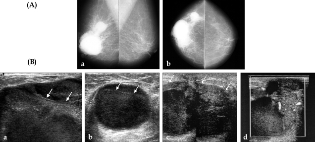
Fig. 1.
(A) Mammogram. Both mediolateral oblique (a) and craniocaudal mammogram (b) showed multiple round shape masses with well-defined margins in the subareolar area and upper outer quadrant of the right breast. (B) Ultrasonogram. The initial sonogram shows well-circumscribed cystic masses with echogenic floating debris (arrows) at the subareolar (a) and upper outer quadrant (b) of the right breast. Sonograms obtained at 5 month follow up. Cystic mass size was markedly increased, with irregular shaped soft tissue (arrows) along the cyst walls (c). The blood flow signals of the papillary solid portion are shown on color Doppler sonography (d).

