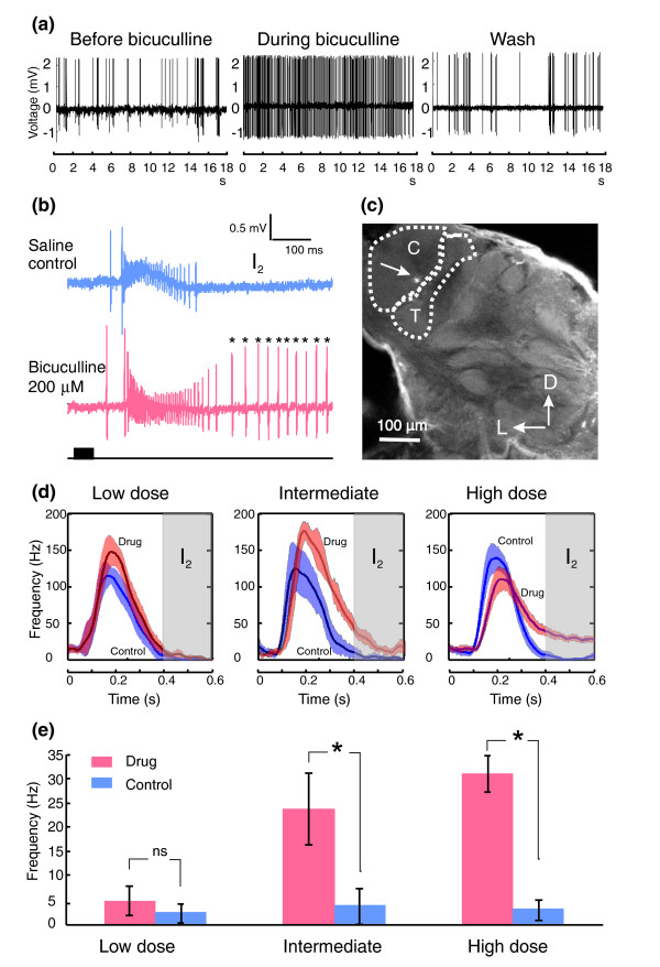
Figure 1.
Effects of bicuculline on the firing pattern of MGC-PNs. (a) Shown as raw spike traces, bath application of 100 μM bicuculline changed the spontaneous firing pattern of an MGC-PN from a random bursting (left) to a more regular tonic pattern (middle). This change was reversed with saline wash (right). (b) The M inhibitory period (I2) that typically follows the odor-evoked excitatory phase in MGC-PNs (upper panel) was completely blocked by treatment with 200 μbicuculline, resulting in an extended excitatory response (asterisks, lower panel). Odor pulse is indicated by the black bar below the traces. (c) Confocal micrograph showing the lucifer yellow fluorescent mark (arrowed) in the cumulus (C) deposited by the glass electrode used to record the C-PN in (b). T, toroid I. (d) Graphs of peristimulus responses (derived from five odor pulses) of 25 MGC-PNs to their specific ligands under saline control (blue curve; mean ± SEM) and bicuculline treatment (orange curve; mean ± SEM) at low (25 μM, n = 8), intermediate (50 μM or 100 μM, n = 7), and high (200–500 μM, n = 10) dosages. The onset of the 50 ms stimulus was at time zero. (e) Histograms derived from the graphs in (d). The shaded areas represent the I2 period, during which the averaged firing rate was not significantly different (NS) between low-dose bicuculline treatment and saline control, but was significantly elevated by intermediate and high-dosage bicuculline treatment. The abbreviation ns and the asterisks respectively indicate non-statistical (Mann Whitney U test, p > 0.05 for low dose, n = 8) and statistical significance (Mann Whitney U test, p < 0.03 for intermediate dose, n = 7; p < 0.001 for high dose, n = 10).

