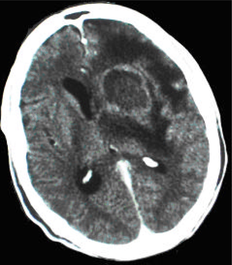
Figure 2.
CT scan (post-contrast) of patient DD showing a nodular, ring-enhancing lesion in the anterior part of the left basal ganglia with accompanying brain edema and moderately raised intracranial pressure. There is a 0.5 mm mid-line shift with lateral ventricle compression on the left. The findings are suggestive of an inflammatory process with abscess formation and a high probability of toxoplasmosis with differential diagnoses of primary CNS lymphoma and a bacterial abscess (less likely mycotic aneurysm, tuberculoma, or metastatic lesion).

