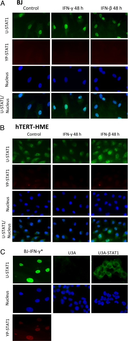
Fig. 4.
Most U-STAT1 is located in nuclei of untreated and IFN-treated BJ or hTERT-HME1 cells. (A) BJ cells were treated with 0.3 ng/mL IFN-γ or 3 units/mL IFN-β for 48 h. Immunocytochemistry was performed with antibodies against U-STAT1 (green) or YP-STAT1 (red). Nuclei were stained with DAPI. YP-STAT1 was not seen in untreated cells or in cells treated with IFNs for 48 h. (B) hTERT-HME1 cells were treated with 0.1 ng/mL IFN-γ or 5 units/mL IFN-β for 48 h. YP-STAT1 was not seen in untreated cells or in cells treated with IFNs for 48 h. (C) As a positive control for PY-STAT1 staining, cells were treated with 3 ng/mL IFN-γ (IFN-γ*) for 1 h, which shows PY-STAT1 staining inside nuclei. U3A cells (STAT1-null 2fTGH cells) were used as a negative control, with same antibodies used in A and B. Untreated U3A cells transfected with wild-type STAT1 show clear cytoplasmic localization of U-STAT. The magnification of the images is ×400.

