
Figure 2.
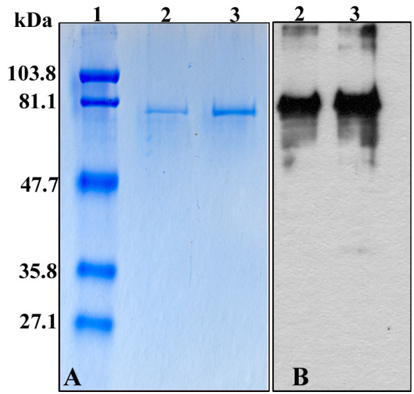
Detection of the purified recombinant Capsid protein 1. Either 0.5 or 1 μg of protein (lanes 2 and 3 respectively) was electrophoresed onto 10% SDS-PAGE and revealed by Coomassie blue staining (A) or Western-blot analysis using anti-Histidine mAb. Standard molecular weight markers (lane 1) are indicated on the left.