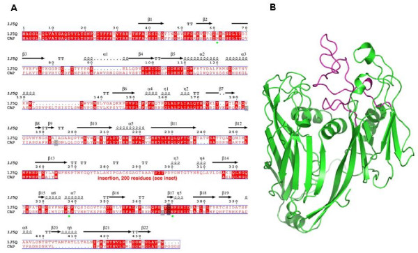
Figure 4.
Sequence alignment and 3D modelling of APM Capsid protein 1 with that of Chlorella virus PBCV1. (A) Sequence numbering is from the Chlorella virus capsid [PDB:1J5Q]. The putative N-glycosylation sites in APM Capsid are identified by green circles. An insertion of 200 AA replaces a coil motif in 1J5Q (positions 265–315) not belonging to the «jelly-roll» motifs. (B) 3D structure representation of Chlorella virus PBCV1 Capsid core domain (green), and the coil domain (magenta) which is substituted in APM Capsid.

