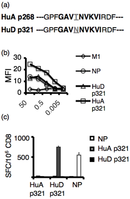
Figure 4. Comparison of HuA p321-specific CD8+ T cells and HuD p321-specific CD8+ T cells.
(a) Sequences of HuD p321 and HuA p321 (b) RMA/S cells were incubated with serial dilutions of peptide and stained for Db MHC I. HuD p321 and HuA p321 were assayed. The A2.1 epitope of influenza (M1) was used as a negative control. The Db epitope of influenza (NP) was used as a positive control. (c) C57BL/6 mice were immunized with individual peptides (NP, HuA p321, or HuD p321) in TiterMax adjuvant (2 mice per group). 7 days later, draining lymph node CD8+ T cells were plated in an IFNγ ELISPOT assay (2×10⁵/well) with peptide pulsed EL4 cells (5×10⁴/well). The assay was performed in triplicate. Means are plotted and error bars represent standard deviations of the mean. Data is representative of four experiments.

