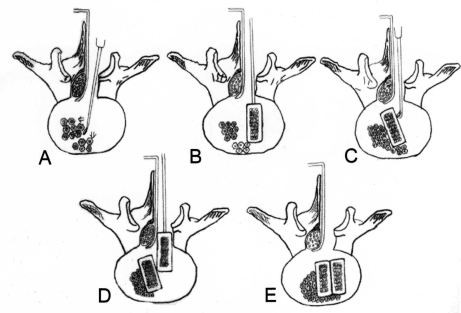
Fig. 1.
Diagrams depicting the steps of the PLIF via a unilateral approach. (A) After the retraction of the thecal sac and traversing nerve root to the midline, disc material and endplates are removed as much as possible in both the contralateral and ipsilateral sides. Before the cage insertion, the lamina and cortical bone from the iliac crest are grafted as much as possible into the contralateral and anterior sides of the intervertebral space. (B) The first cage filled with cancellous bone from the iliac crest is introduced to the intervertebral space. (C) The cage is carefully pushed to the contralateral side with the down-biting curettes and impactor. (D) The second cage was impacted to the ipsilateral side in the same manner. (E) Lastly, adequate impaction and complete hemostasis are performed.

