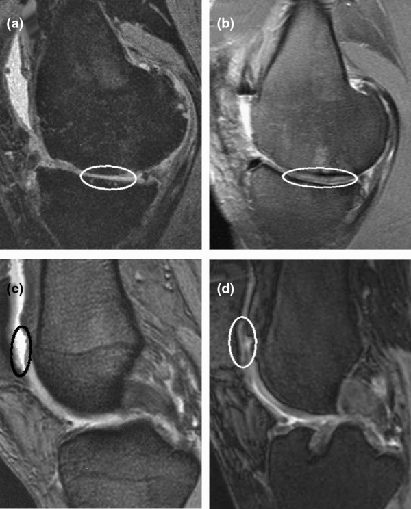
Figure 7.

Magnetic resonance imaging (MRI) before and four years after implantation of BioSeed®-C. (a) Preoperative MRI shows a cartilage defect (encircled) at the medial femoral condyle. (b) After four years, MRI documented complete filling of the defect. Preoperatively, MRI shows a patellar (c) cartilage defect (encircled) that was completely filled after implantation of the graft as assessed by MRI at four years (d).
