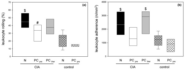
Figure 3.
Primary and secondary leukocyte–endothelial cell interactions in synovial venules of animals with collagen-induced arthritis. Quantitative analysis of (a) primary (rolling) and (b) secondary (firm adherence) leukocyte–endothelial cell interactions in the synovial venules of animals with collagen-induced arthritis (CIA) and either the normal diet (N) or the phosphatidylcholine-enriched diet, starting either with the CIA induction (PCpre) or with the clinical onset of the disease (PCther). For the induction of CIA, animals were immunized twice with collagen II and complete Freund's adjuvant/incomplete Freund's adjuvant. Three weeks after the second immunization, the knee joints were assessed by intravital fluorescence microscopy, as described in Materials and methods. Values given as medians with the 25th and 75th percentiles. #P < 0.05 versus N/CIA. $P < 0.05 versus N/control.

