
Figure 1.
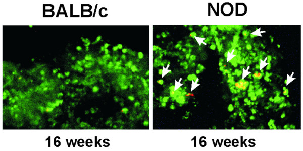
Acinar cell suspension. Acinar cells were stained with acridine orange and propidium iodide (AO/PI). Nuclei of live cells fluoresce green with AO/PI under fluorescence microscopy. Arrows indicate apoptotic cells with yellow-orange fluorescence. These images are representative of five others from similar experiments run with independent non obese diabetic (NOD) and BALB/c acinar samples. Magnification ×200.