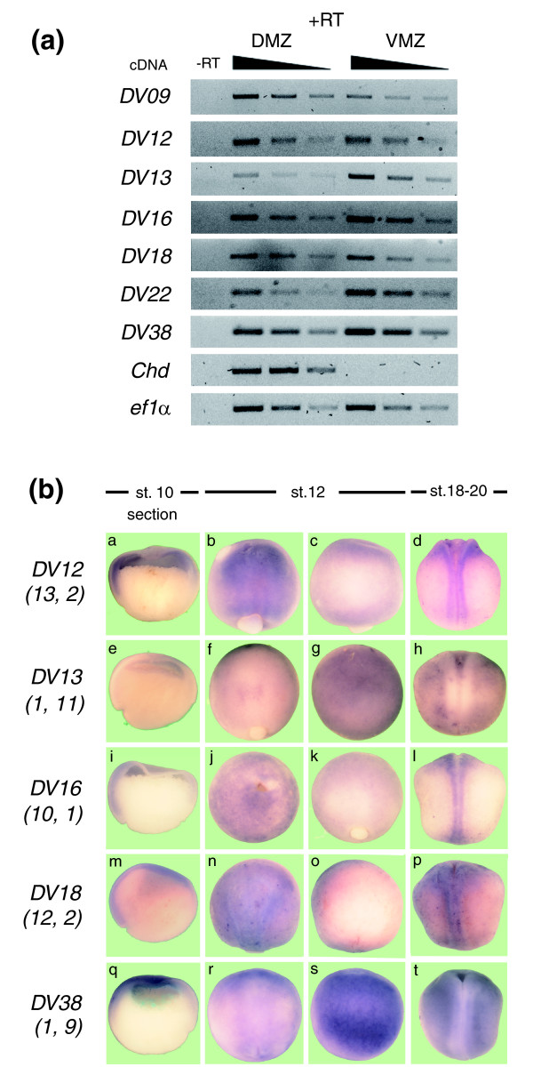
Figure 4.
Verification of the differential expression of X. laevis homologues. (a) Total RNA was isolated from dorsal (DMZ) and ventral (VMZ) explants at the gastrula stage. RT-PCR was performed using specific primers for each transcript and different cDNA concentrations (serial dilutions of cDNA, 1:1, 1:2 and 1:4). Chordin was included as control. Reverse transcription in the absence (-RT) or presence (+RT) of reverse transcriptase for specificity of cDNA amplification. (b) X. laevis embryos at stage (st.) 10 (a, e, i, m, q; hemi-sections, dorsal to the left), stage 12 (b, c, f, g, j, k, n, o, r, s; anterior is up) and stages 18-20 (d, h, l, p, t; anterior is up) were processed for in situ hybridization with specific probes for each transcript. Stage 12 embryos are pictured from both sides relative to the blastopore to illustrate its asymmetric expression. Numbers under each transcript correspond to the frequency of occurrence in each SAGE library (tag frequency in dorsal library; tag frequency in ventral library).

