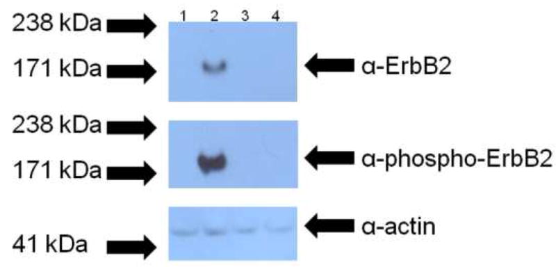
Figure 5.

Western blot analysis of ErbB2 activity in stable pools of CHO and CHO-bcl-xL cells. Samples containing equal amounts of total cellular protein from stable pools of CHO and CHO-bcl-xL cells transfected with pcDNA3.1/zeo_erbB2 (lanes 1 and 2, respectively) and transfected with the empty vector (lanes 3 and 4, respectively) were analyzed to determine if the expressed ErbB2 was functional. The top panel shows the expression of ErbB2, and the middle panel shows the activity of the protein as assessed by the receptor’s phosphorylation. The bottom panel shows the corresponding anti-actin blot to ensure that equal cellular protein was loaded in each lane. All samples were run on the same Western blot. The arrows on the left indicate where the corresponding molecular weight marker from the protein ladder ran.
