
Figure 1. Effect of Cavβ subunits on cluster organization of Cav1.2 channels.
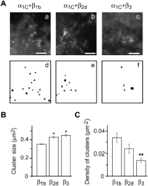
(A) TIRF images (a–c) and wavelet-derived clusters (d–f) of Cav1.2 channels containing β1b (a,d), β2d (b,e) or β3 (c,f). Scale, 4.5 µm. (B) Dependence of the average size of Cav1.2 clusters on the type of Cavβ present. β1b, mean size±SEM, 0.360±0.005 µm2 (number of clusters analyzed m = 1253); β2d, 0.430±0.013 mm2 (m = 270); β3, 0.450±0.017 µm2 (m = 205). *, P<0.001 relative to β1b. (C) Dependence of the number of Cav1.2 clusters (normalized to the area measured and defined as density) on the type of Cavβ present. β1b, mean number±SEM, 0.034±0.004 mm−2 (number of cells n = 27); β2d, 0.024±0.04 µm−2 (n = 30); β3, 0.014±0.02 µm−2 (n = 22). **, P<0.01 relative to β1b. Vα1C was co-expressed with α2δ and indicated Cavβ in COS1 cells.