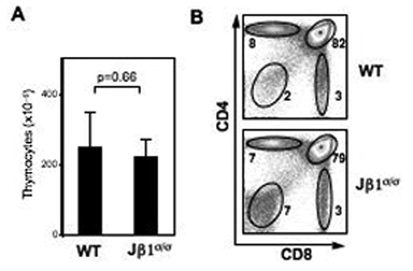
Figure 2. Thymocyte development in Jβ1σ/σ mice.

A. Numbers of total thymocytes in Jβ1σ/σ (n=4) and wild-type (WT) (n=4) mice. B. Flow cytometric analysis of thymocyte development, using anti-CD4 and anti-CD8, in WT and Jβ1σ/σ mice. Percentages are indicated beside each gate. Results are representative of at least 3 independent experiments.
