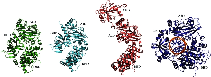
Figure 2.
Different conformations of archaeal and human ATP-dependent DNA ligases. Ribbon diagrams of A. fulgidus DNA ligase, P. furiosus DNA ligase, S. solfataricus DNA ligase and human DNA ligase I fragment (with bound DNA) are drawn in green, cyan, pink and blue, respectively. A. fulgidus DNA ligase shows the most closed conformation, with water-mediated hydrogen-bond interactions between the two terminal domains DBD and OBD.

