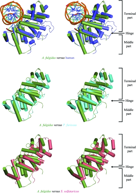
Figure 4.
Superposition of the middle part of the DBD in four DNA ligases in stereo. The DBD of A. fulgidus DNA ligase, human DNA ligase I fragment (with bound DNA), P. furiosus DNA ligase and S. solfataricus DNA ligase are coloured green, blue, cyan and pink, respectively. The superposition shows that the conformation of DBD in A. fulgidus DNA ligase is more similar to that of the human DNA ligase I fragment than to those of the P. furiosus and S. solfataricus DNA ligases.

