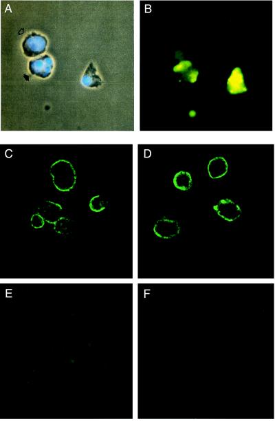
Figure 3.
Immunofluorescence microscopy of apoptosis-induced PAECs. (A) DNA staining with Hoechst dye showed normal cell with single nucleus (open arrow) and apoptotic cell with characteristic fragmented nuclei (closed arrow). Note apoptotic body below. (B) Binding of EO6 to the cells in the same field observed under a fluorescence microscope. The upper normal cell showed diffuse nonspecific fluorescence, whereas the apoptotic cells and body showed specific punctuate fluorescence. Confocal microscopy of apoptosis-induced PAECs: (C) EO6 and (D) EO14 bound to surface of apoptotic cells but not to (E) normal PAEC. (F) Apoptotic PAEC stained with control mouse IgM showed nonspecific fluorescence.

