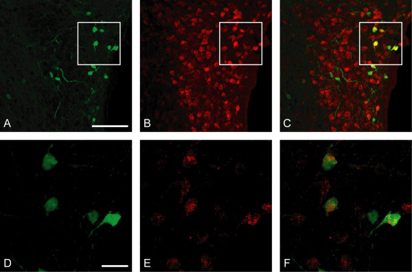
Figure 2.
Expression of EGFP/connexin 36 in a subset of Fluorogold-labeled neuroendocrine cells in PVN. Confocal photomicrographs show immunofluorescence for EGFP (A, D; green), Fluorogold (B, E; red), and their colocalization (C; yellow) for A, B and (F; yellow) for D, E. Photomicrographs D-F show neurons from insets in A-C in higher magnification. The third ventricle is seen to the right in A-C. Scale bars = 100 μm in A-C and 20 μm in D-F.

