Figure 1.
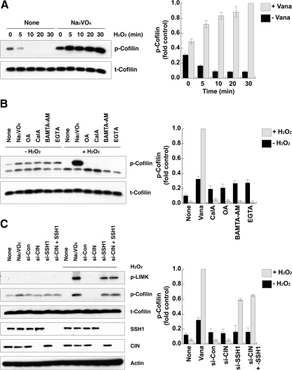
SSH-1L participates in H2O2-induced cofilin dephosphorylation. (A) HeLa cells were pretreated with or without 0.1 mM Na3VO4 for 30 min and then stimulated with 0.5 mM H2O2 for the indicated times. Cell lysates were immunoblotted with antibodies to phosphorylated cofilin (p-Cofilin) or cofilin (t-Cofilin). (B) HeLa cells were pretreated with or without the indicated inhibitor sets (1 mM Na3VO4, 1 μM okadaic acid [OA], 1 μM CalyculinA [CalA], 30 μM BAMTA-AM, 1 mM EGTA) for 0.5 h and then stimulated with 0.5 mM H2O2 for 30 min. Cell lysates were immunoblotted with antibodies to phosphorylated cofilin (p-Cofilin) or cofilin (t-Cofilin). (C) HeLa cells were transfected with 10 nM control, CIN or SSH-1L siRNA (si) for 48 h and then stimulated with or without 0.5 mM H2O2 for 30 min. Cell lysates were immunoblotted with antibodies to phosphorylated LIMK1/2 (p-LIMK), phosphorylated cofilin (p-Cofilin), cofilin (t-Cofilin), SSH-1L, CIN, or actin. (A–C) The graphs at the right of each panel represent the averaged normalized p-cofilin values; the value in Na3VO4 + H2O2-treated cells is taken as 1.0. Values are from three independent experiments ± SD.

