Figure 3.
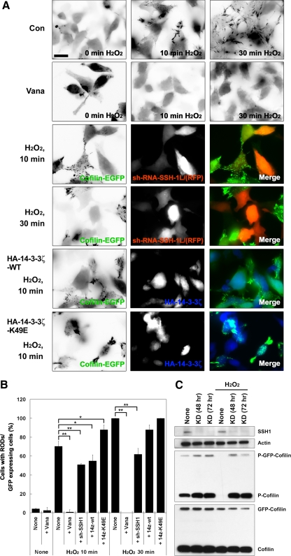
H2O2 induces cofilin rod formation in cofilin-GFP–expressing HeLa cells. (A) Cofilin-GFP–expressing HeLa cells transfected with or without sh-RNA-SSH-1L/RFP, 14-3-3ζ-wt, or 14-3-3ζ-K49E were treated with or without H2O2 for up to 30 min, as indicated. The eGFP-cofilin images are presented as inverted fluorescence images to highlight rod formation. The merged images show eGFP-Cofilin (green), sh-RNA-SSH-1L/RFP (red), or HA-14-3-3ζ (blue). Scale bar, 10 μm. (B) Cofilin rod formation is quantified and represented graphically. The percentage of rod-forming cofilin-GFP–expressing HeLa cells was scored as the total number of rod-containing cells/total GFP-positive cells. The number of rods observed in H2O2-treated cells at 30 min was set to 100%. The average ± SD of three experiments is shown, where a minimum of 100 cells were scored for each time point (* p > 0.05; ** p > 0.01). (C) Protein expression level of the cells treated with sh-RFP-SSH-1L was confirmed using Western blotting with anti-SSH-1L, -actin, -phosphorylated cofilin, or -cofilin antibodies. Knockdown (KD) of SSH-1L prevented the H2O2-induced loss of p-Cofilin and p-GFP-Cofilin. Representative experiments from three separate experiments are shown.

