Figure 6.
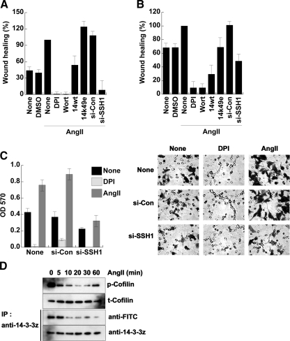
AngII-induced cell migration is dependent on ROS and SSH-1L. (A and B) Confluent HeLa (A) or MCF-7 (B) cells seeded onto 60-mm culture dishes were wounded, washed twice with PBS, and treated with or without DMSO or 0.1 μg/ml AngII, as described in Materials and Methods. Phase-contrast images were taken 24 h after wounding and treatment, and the number of cells migrating into the wounded area was determined using ImageJ software. Cells were pretreated with DPI (10 μM) or Wort (100 nM) as indicated for 30 min before AngII stimulation. Another set of cells was transfected with 14-3-3ζ-wt, -K49E, siRNA-con, or siRNA-SSH-1L for 48 h before seeding onto dishes. 100% of wound healing is defined for AngII-stimulated cells. (C) MCF-7 cells were transfected with or without siRNA (si)-control or siRNA-SSH-1L RNA for 48 h and then transwell migration assays were performed as described in Materials and Methods. (A–C) Averages ± SD or n = 3 experiments. (D) MCF-7 cells were stimulated with 0.1 μg/ml AngII for the indicated times. Cell lysates were immunoblotted with antibodies to phosphorylated cofilin (p-Cofilin) or cofilin (t-Cofilin) and labeled with 5′-iodoacetamide fluorescein, as in Materials and Methods. The labeled proteins were immunoprecipitated (IP) with anti-14-3-3ζ antibodies, followed by immunoblotting with anti-FITC antibodies. The total immnoprecipitated proteins were analyzed by immunoblotting with anti-14-3-3ζ antibody. Loss of anti-FITC reactivity is indicative of protein oxidation. A representative experiment from two separate experiments is shown.

