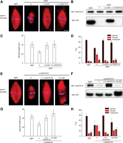
Figure 1.
Nonredundant roles of EB1 and XMAP215 in the regulation of spindle length and bipolarity. (A) Representative images of fixed metaphase-arrested spindles assembled around replicated sperm in control depleted (ΔIgG) or EB1 depleted (ΔEB1) extracts supplemented with either recombinant EB1 (wild-type levels, ΔEB1 + 1× EB1) or a onefold excess of XMAP215 (ΔEB1 + 1× XMAP215). Microtubules (red) are visualized by Cy3-tubulin and DNA (blue) is stained with Hoechst. Bar, 10 μm. (B) Accompanying immunoblots of extracts shown in A. (C) Average length of sperm-spindles (conditions as indicated in A). Error bars represent SD (n = 50 bipolar spindles per condition; one representative experiment is shown). (D) Quantification of pole numbers in the spindles assembled around sperm nuclei in extracts treated as described in A. Error bars represent SD (n = 100 structures per condition; average of two independent experiments). (E) Representative images of fixed metaphase-arrested spindles assembled around replicated sperm in control depleted (ΔIgG) or ∼70% depleted XMAP215 (∼ΔXMAP215) extracts supplemented either with recombinant XMAP215 (wild-type levels, ∼ΔXMAP215 + 1× XMAP215) or a threefold excess EB1 (∼ΔXMAP215 + 3× EB1). Microtubules (red) are visualized by incorporated Cy3-tubulin and DNA (blue) is stained with Hoechst. Bar, 10 μm. (F) Accompanying immunoblots of extracts shown in E. (G) Average length of sperm-spindles under the conditions described in E. Error bars represent SD (n = 50 bipolar spindles per condition; one representative experiment is shown). (H) Quantification of pole numbers in spindles formed around sperm nuclei in extracts treated as described in E. Error bars represent SD (n = 100 structures per condition; average of two independent experiments).

