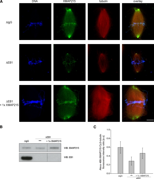
Figure 4.
EB1 increases binding of XMAP215 to the spindle microtubules. (A) Representative, maximum intensity Z-projections of fixed spindles assembled around sperm nuclei in cycled metaphase-arrested extracts that were control-depleted (ΔIgG), EB1-depleted (ΔEB1), or EB1-depleted and supplemented with a onefold excess of XMAP215 (ΔEB1 + 1× XMAP215). Microtubules are red (Cy3-tubulin), DNA is blue (Hoechst), and XMAP215 is shown in green (indirect immunofluorescence). Bar, 10 μm. (B) Immunoblots of extracts treated as described above and probed with anti-XMAP215 and anti-EB1 antibody. (C) Average ratio of XMAP215 indirect fluorescence intensity and Cy3-tubulin fluorescence intensity calculated from maximum intensity Z-projections of spindles treated as described above. Error bars represent SD (n = 30 spindles per condition; one representative experiment).

