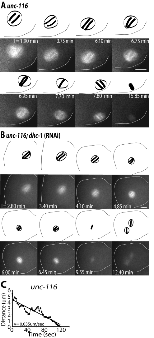
Figure 2.
unc-116;dhc-1(RNAi) spindles fail to undergo directed movement toward the cortex. Images of GFP-tubulin fluorescence within a meiotic embryo are shown from representative time-lapse sequences from an unc-116 worm (A) and a unc-116;dhc-1(RNAi) worm (B). Zero minutes (0 min) corresponds to exit of the embryo from the spermatheca. The cell cortex was highlighted in each image for clarity. Drawings corresponding to each image are included for clarity to show spindle orientation and length. Spindle orientation was determined from dense bundles of microtubules that extend along the pole-to-pole axis. (A) The unc-116 spindle failed to translocate early to the cortex and remained stationary at a position several micrometers from the cortex from 0 to 6.70 min. Spindle shortening initiated just before the start of late translocation to the cortex (6.75–7.80 min). The spindle undergoes late, pole-leading translocation to the cortex ending perpendicular to the cortex (7.80 min). (B) In the unc-116;dhc-1(RNAi) embryo, the spindle remained away from the cortex throughout meiosis and failed to undergo directed movement toward the cortex. The spindle completed meiosis I away from the cortex and two meiosis II spindles assembled (T = 12.4 min). Corresponding Supplemental Videos 3 and 4 can be found in the online supplemental material. (C) Representative graph of unc-116 spindle movement over time after spindle shortening. Distance was measured as described for wild type in Figure 1 (C). The unc-116 spindle translocated to the cortex following a linear track with a velocity of 0.035 μm/s. Bars, 5 μm.

