Figure 1.
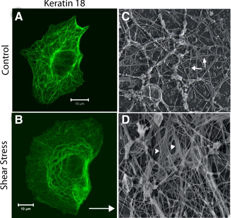
Shear stress results in the structural reorganization of the KIF network. A549 cells exposed to static control and shear stress (30 dyn/cm2; 60 min; arrow in B indicates direction of flow) were fixed and processed for indirect immunofluorescence using anti-K18. In cells exposed shear stress the KIF network was reorganized into thick, wavy tonofibrils; representative photomicrographs are shown in A and B. Cells were grown on glass coverslips and exposed to shear stress (30 dyn/cm2; 4 h). The cells were extracted with a buffer containing 100 mM PIPES, pH 6.9, 1 mM MgCl2, 1 mM EDTA, 0.4 M NaCl, 1% TX-100, 0.5 mg/ml DNase I, and 4% polyethylene glycol for ∼4 min at RT, then treated with the N-terminal domain of gelsolin to remove actin, fixed, dehydrated, and dried by the critical point method. These preparations were also devoid of microtubules. Once dried, the preparations were rotary shadowed with carbon/platinum, and the shadowed replicas were transferred to copper grids for observation by transmission electron microscopy (for details see Helfand et al., 2002) Representative electron micrographs show that the KIF network is comprised of individual ∼10-nm filaments in control cells (Figure 1C); after shear stress the formation of tonofibrils comprised of bundles of KIF was observed (Figure 1D; arrowheads in D indicate formation of keratin “tonofibrils”). White scale bar, 10 μm.

