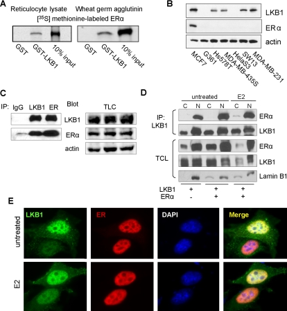
Figure 1.
LKB1 binds to ERα. (A) Left, GST pull-down assays were conducted using 35S-ERα incubated with GST and GST-LKB1 (50 pmol) fusion proteins and visualized by autoradiography. (B) Western blot analysis of LKB1 and ERα expression in different cancer cell lines was confirmed by western blot analysis using antibodies specific for LKB1, ERα, and actin. (C) Anti-LKB1, -ERα, and IgG (control) antibodies were used to IP endogenous proteins from MCF7 cells, followed by Western blot analysis for IP LKB1, ERα, and TCL in which actin was used as loading control. (D) MCF cells were transfected with pcDNA3.1, ERα and LKB1 expression plasmids. Cells were treated with E2 (100 nM) followed by cellular fractionation: nuclear (N) and cytoplasmic (C). LKB1 was IP using anti-LKB1 antibody, followed by Western blot analysis in which membranes were probed for LKB1, ERα, and lamin B1 (nuclear protein marker). (E) Representative immunofluorescent images of MCF7 cells left untreated or treated with E2 (100 nM), fixed, and incubated with anti-LKB1 (green) and anti-ERα (red) antibodies and appropriate secondary antibodies. Nuclei were stained with DAPI. Results are representative of three experiments.

