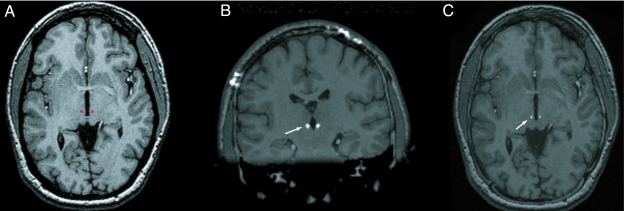
FIGURE 2.
A, Preoperative magnetic resonance imaging with a surgical plan of a patient with Tourette syndrome, in which the centre médian/parafascicular thalamic nucleus was targeted (red dots). Coronal (B) and axial (C) postoperative computed tomogram fused with preoperative magnetic resonance imaging showing Medtronic 3387 lead in the centre médian/parafascicular nucleus of thalamus (white arrows).

