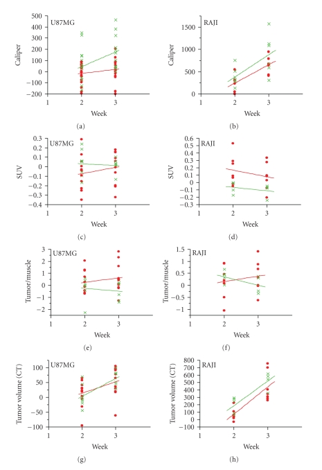
Figure 2.
Tumor assessments of Xenografts U87MG and RAJI at 2 and 3 weeks after enzastaurin treatment. Standard caliper measurements show a tumor growth delay for U87MG (P < .05 at week 3), but not for RAJI (Panels (a) and (b)). [18F]FDG-PET imaging was performed at the same time as standard caliper measurements (Panels (c)-(d)). Tumor glucose metabolism changes were measured by SUV (Panels (c), (d)) and tumor/muscle ratio (Panels (e), (f)) in U87MG and RAJI xenografts. Only in RAJI xenografts enzastaurin treatment showed a trend in increased SUV (Panel (d); P < .10 at weeks 2 and 3). Using tumor/muscle ratio indicated a trend for FDG uptake in U87MG (P < .10 at week 3) (Panel (e)), but not in RAJI xenografts (Panel (f)). The tumor size assessment based on CT scan (Panels (g) and (h)) indicated a trend for detecting reduced tumor size only in RAJI xenografts after enzastaurin treatment at week 2 (P < .10). Mice treated with vehicle alone are shown in green; mice treated with enzastaurin are shown in red, representation of 2 independent experiments.

