
Fig. 1.
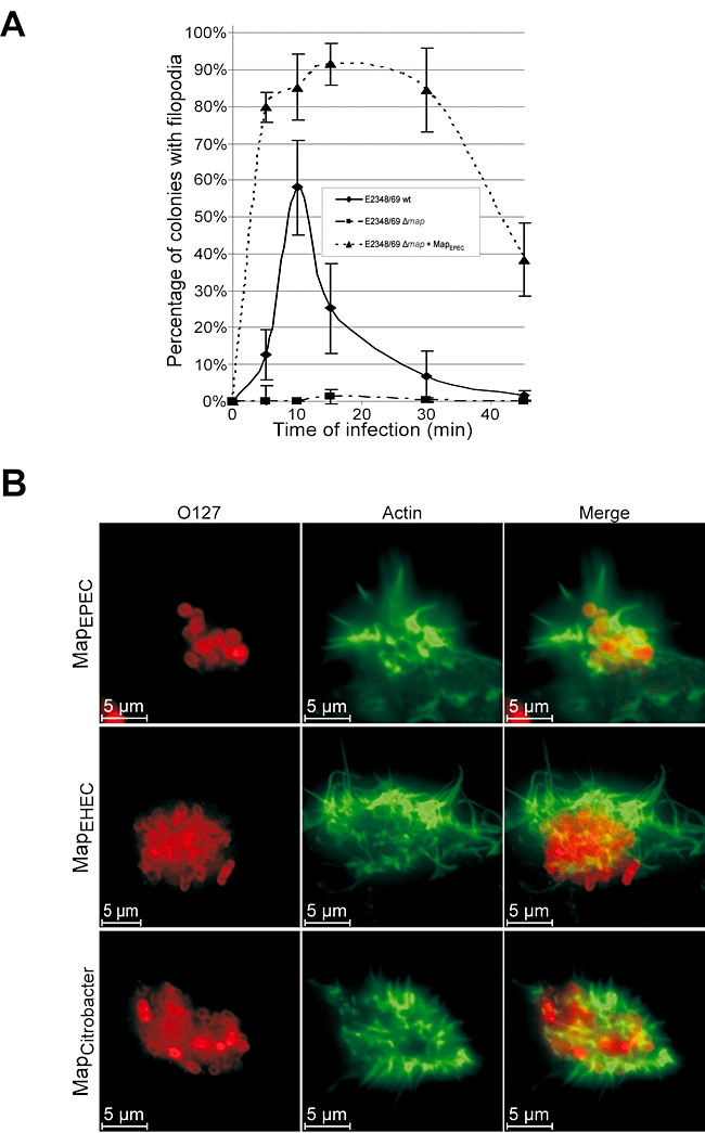
Kinetic of filopodia formation on 3T3 Swiss cells. A. Quantification of microcolony-associated filopodia on cell infected with wild-type E2348/69, E2348/69Δmap and E2348/69Δmap overexpressing MapEPEC. One hundred cells were counted in five independent experiments. Results are presented as mean ± SD. B. Fluorescence microscopy of 3T3 cells infected for 15 min with E2348/69Δmap overexpressing Map from EPEC, EHEC or C. rodentium. Actin was stained with Oregon green phalloidin (Green) and EPEC were detected with rabbit anti-0127 antibody (Red).