
Fig. 2.
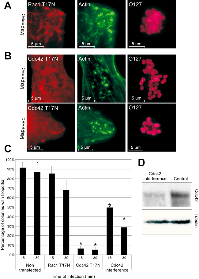
Filpodia formation by Map is Cdc42-dependent. 3T3 cells were transfected with dominant-negative Rac-1T17N (A) or Cdc42T17N (B) 24 h prior to infection with E2348/69Δmap overexpressing MapEPEC or MapEHEC for 15 min. Actin was stained with Oregon green phalloidin (Green), the Myc-tagged GTPases with mouse anti-myc (Red) and EPEC with rabbit anti-O127 (Magenta). Filopodia are observed on cells transfected with dominant-negative Rac-1 (A), but not on cells transfected with dominant-negative Cdc42 (B). Quantification of microcolony associated with filopodia in 3T3 cells transfected with dominant-negative Cdc42 or Rac-1 and Cdc42 siRNA (C). Cells were infected for 15 or 30 min with E2348/69Δmap overexpressing MapEPEC. One hundred colonies on transfected cell were counted in five independent experiments. Results are presented as mean ± SD. Significant differences from non-transfected cells are indicated by asterisks (*P < 0.01). Presence of filopodia induced by Map is affected by expression of Cdc42T17N or Cdc42 siRNA, but not by expression of Rac-1T17N. The level of Cdc42 and Tubulin in cell lysates 48 h after transfection with Cdc42 siRNA was determined by Western blots (D).