
Fig. 3.
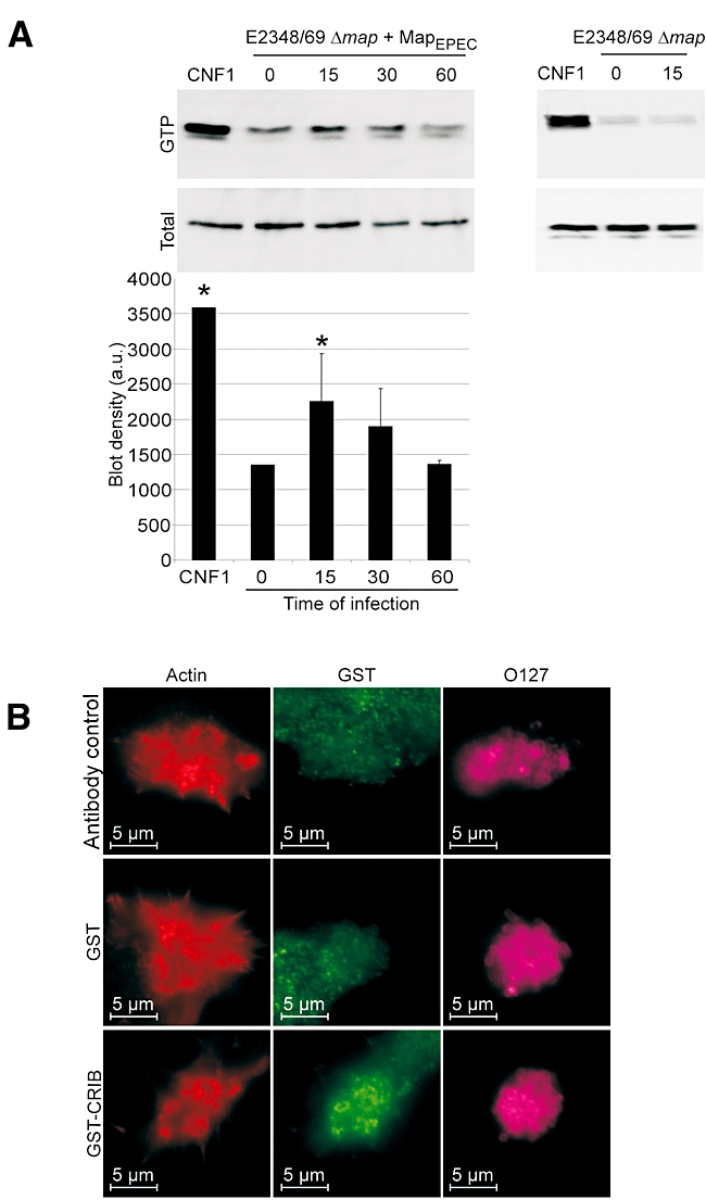
Map activates Cdc42. A. Swiss 3T3 cells were infected with E2348/69Δmap overexpressing MapEPEC for 15, 30 or 60 min. CNF1 toxin was used as a positive control (25 μg ml−1). Cells were lysed and a GST–CRIB fusion protein was used to co-purify Cdc42-GTP. Total Cdc42 in the lysates and Cdc42-GTP were detected by Western blotting with anti-Cdc42 antibodies. The graph shows measurement of blot densities from three pull-down experiments (means ± SD). Significant differences from uninfected cells (time 0) are indicated by asterisks (*P < 0.05). Infection with E2348/69Δmap did not induce Cdc42 activation. B. Fluorescence microscopy of 3T3 cells infected for 15 min with E2348/69Δmap overexpressing MapEPEC. Fixed cells were pre-incubated with purified GST or GST–CRIB. Actin was stained with TRITC phalloidin (Red), GST and GST–CRIB were detected with rabbit anti-GST antibody (Green) and EPEC were detected with goat anti-E. coli antibody (Magenta). Specific signals, at the base of filopodia, were observed in cell treated with GST–CRIB whereas no signal could be detected for control antibody or GST alone.