Fig. 1.

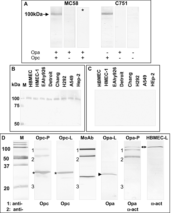
Direct interactions of Opc-expressing Nm variants with a 100 kDa protein of human endothelial and epithelial cells. A. Proteins from whole-cell lysates of HUVEC were separated by SDS-PAGE and Western blotted. Blots were overlaid with Nm variants either expressing or lacking the expression of the opacity proteins Opa and Opc as shown. After 2 h, the binding of bacteria was detected with rabbit antiserum against Nm followed by AP-conjugated anti-rabbit antibody. Strip marked with asterisk (*) represents a control that excluded anti-Nm primary antibody.
B and C. Whole-cell lysates of a number of diverse epithelial and endothelial cell lines were solubilized in octylglucoside and subjected to SDS-PAGE and Western blotted. Blots were overlaid with Opc+(B) or Opc− (C) variants of strain C751 and bacterial binding detected as in (A). Lanes marked M contained molecular weight markers. Data are from one representative of two independent experiments. D. Co-precipitation of proteins from whole-cell lysates of bacteria and target cells. Whole-cell lysates of Nm C751 isolates Opc+(Opa−) or Opa+(Opc−) and HBMEC were mixed and any α-actinin–ligand complexes were pulled down using the monoclonal antibody 7H6 against α-actinin. The complex was dissociated and subjected to SDS-PAGE and Western blotted. The lanes marked Opc-P and Opa-P contained the co-precipitated samples from HBMEC incubated with Opc- or Opa-expressing Nm. Controls included lanes loaded with the mouse mAb 7H6 alone (lane marked MoAb), whole-cell lysates of Opc+Opa− or Opc−Opa+ bacteria (lanes marked Opc-L and Opa-L respectively) and whole-cell lysates of HBMEC (lane marked HBMEC-L). The blots were developed using primary antibodies as shown and appropriate alkaline phosphatase-conjugated secondary antibodies. In some cases, the blots were sequentially developed with two antibodies. The bands marked 1, 2 and 3 are derived from the anti-α-actinin mAb 7H6. Band 3, presumably the light chain of the antibody, was often faintly stained. Asterisks show the position of Opc protein in bacterial lysates (Opc-L) and in the lane Opc-P containing the co-precipitated proteins pulled down by the anti-α-actinin antibody 7H6. Whereas the Opa protein (arrowhead in lane Opa-L) was not co-precipitated as seen in Opa-P lane, which was also developed using α-actinin antibody to show that the precipitate did contain α-actinin (arrow). Lanes marked M contained molecular weight markers.
