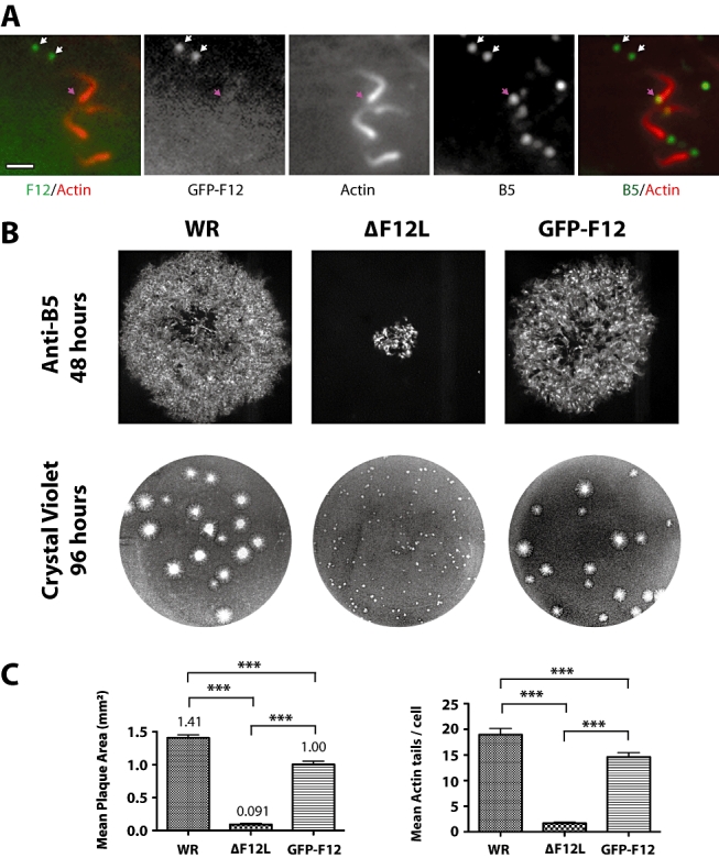
Fig. 2.

GFP-F12 rescues actin tail formation and cell-to-cell spread of ΔF12L virus. A. Immunofluorescence images of WR-GFP-F12-infected HeLa cells reveals that GFP-F12 is associated with B5-positive IEV particles (white arrows) but is absent from the tips of actin tails (pink arrow). Scale bar = 2 μm. B. Representative images of plaques formed by WR, ΔF12L and WR-GFP-F12 in confluent BS- C-1 monolayers at 48 (anti-B5) and 96 h (crystal violet) post infection. C. Quantitative analysis of plaque size at 48 h post infection and the number of actin tails per cell in WR, ΔF12L and WR-GFP-F12 8 h post infection. Error bars represent the SEM and were derived from measuring the area of 20 plaques or counting 150 infected HeLa cells. ***P < 0.001.
