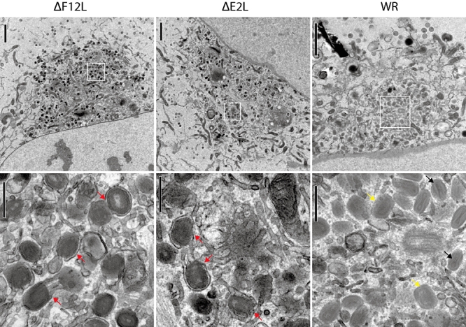
Fig. 10.

E2 and F12 are required for IEV morphogenesis. Electron micrographs showing the peri-nuclear wrapping compartment in WR-, ΔE2L- and ΔF12L-infected HeLa cells at 8 h post infection. In WR-infected cells IMV (black arrows) and wrapped brick-shaped IEV (yellow arrows) are readily observed. In contrast, in ΔE2L- and ΔF12L-infected cells, IEV are partially wrapped (red arrows) and embedded in a dense tubular network of membranes. Scale bars = 2 μm top panels and 0.5 μm bottom panels.
