
Figure 3. βTRCP targets REST during oncogenic transformation.
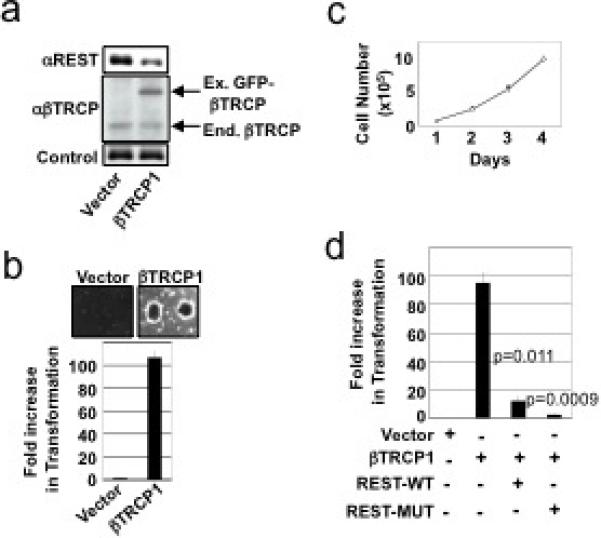
a, TLM-HMECs were transduced with control or GFP-βTRCP1-expressing retroviruses. Lysates were probed with antibodies against REST (upper panel), βTRCP (middle panel), or Vinculin (lower panel). b, Cells from a were analyzed for anchorage-independent colony formation. Assays were performed in quadruplicate (error bars +/− s.d.). Representative of 3 independent experiments is shown. c, HMECs were transduced with retroviruses expressing wild-type REST (REST-WT), degron-mutant REST (REST-MUT), and/or βTRCP1. Cell numbers were monitored for 4 days after plating on tissue-culture dishes. (open circles: vector-1+vector-2, closed circles: βTRCP1, open triangles: βTRCP1+vector-2, closed diamonds: βTRCP1+REST-WT, open squares: βTRCP1+REST-MUT) d, Cells from c were assessed for anchorage-independent colony formation. Assays were performed in triplicate (error bars +/− s.d.). Representative of 2 independent experiments is shown.