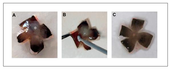
Figure 4.
The human fetal eye is dissected into 4 quadrants. An incision should be made as close to the optic disc with a razor blade in order for the eye cup to lay flat (a). The sensory retina (asterisk) is firmly attached to the optic disc. The sensory retina is gently removed from the RPE layer with a sterile disposable inoculating loop, care being taken not to make scratches into the RPE layer (b). After removal of retina, there is no obvious damage to the RPE layer (c).

