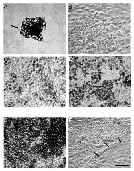
Figure 8.
Light microscopic images showing pattern of time dependent growth of normal RPE in a T75 flask (a-d), and highly pigmented (e) and overgrown multi-layers of RPE (f) for comparison. (a) Day1; Cells are attached and initial growth of the culture occurred in the form of small islands, consisting of 15-25 RPE cells. (b) Day4; Rapid growth of cells with a decrease in pigment is seen. (c) Day14; Cells show evidence of repigmentation. (d) Day21; Cells showing hexagonal shape and increase in pigment density. (e) Hyperpigmented RPE on day 21. These cell are not suitable for establishing polarity. (f) RPE cells with abnormal morphology on day 21. Arrows show cells with fibroblastic morphology. Bar equals 60 μm.

