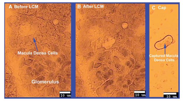
Fig 1. Isolating macula densa cells with laser capture microdissection (LCM).
A. Macula densa cells were identified by their anatomical location and morphology with the LCM microscope in a frozen slide of kidney cortex from SD rat. B. Macula densa were captured with LCM, using a beam width of 7.5 μm and a beam intensity of 50 mW. C. Captured macula densa cells on the cap.

