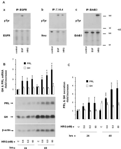
Fig. 1. Effects of heregulin on PRL and GH.
A) ErbB receptor expression in GH4C1 cells: GH4C1 cells were serum-starved overnight, treated with EGF (5 nM) or HRG (6 nM) for 10 min and total protein extracted. Immunoprecipitations (IP) were performed with (Aa) EGFR (1005), (Ab) p185 (Ab 7.16.4) and (Ac) ErbB3 (C-17) antibodies as indicated. Immunoblotting (IB) was performed with pTyr (PY99) antibody (upper panels). Subsequently, membranes were stripped and re-blotted with EGFR (1005), Neu (C-18) and ErbB3 (C-17) antibodies (lower panels).
B-C) Time and dose-dependent effects of heregulin on PRL and GH expression: GH4C1 cells were serum-starved for 24 hrs and subsequently treated with indicated HRG concentrations for up to 48 hrs. B) Total RNA was extracted at the times indicated and GH mRNA expression determined by Northern blot. Subsequently, membranes were stripped and reblotted with specific probes for PRL and β-actin, respectively. The ratio of GH or PRL mRNA vs. β-actin mRNA was calculated by densitometric analysis of each treatment group. The GH / β-actin ratio and PRL / β-actin ratio of the control group at 24 hrs was set as 1.0, and relative mRNA expression levels normalized (mean ± SE; upper panel). Results shown are representative of three independently performed experiments (lower panel). C) GH and PRL secretion in the culture medium were determined by RIA. GH and PRL secretion levels of the control group at 24 hrs were set as 1, and relative secretion levels normalized (mean ± SE of three independently performed experiments). *, p<0.05; **, p<0.01; ***, p<0.001.

