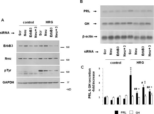
Fig. 4. Pituitary p185c-neu / ErbB3 heterodimer signaling.

GH4C1 cells were serum-starved overnight, transfected with negative control (Scr), p185c-neu (Neu), ErbB3 (100 pmol) or both p185c-neu and ErbB3 (50 pmol each) siRNA (24 hrs). After an additional 8 hr (serum-free medium), cells were treated with HRG (6 nM) for 10 min (A) or 48 hrs (B - C). Expression levels of ErbB3, p185c-neu, pTyr and GAPDH loading control as determined by Western blot of whole cell extracts are depicted (A). GH and PRL mRNA expression and protein secretion were determined as for Fig. 1. Representative of 3 independently performed experiments is shown (mean ± SE). *, p<0.05; **, p<0.01 vs. Scr control group; #, p<0.05; ##, p<0.01 vs. Scr HRG group.
