
Figure 7. TNFα increases the amplitude and frequency of focal calcium bursts by a nSMase2-dependent mechanism.
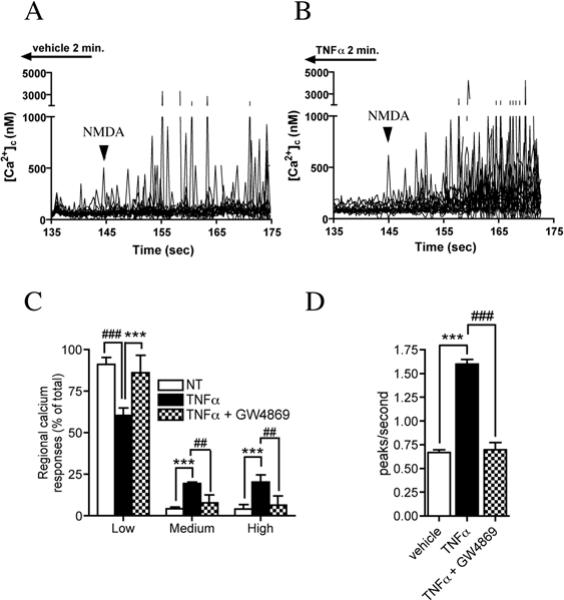
Calcium flux was measured in ∼1.0 μm2 regions along the dendrites of hippocampal neurons in culture using the calcium binding dye Fura-2FF. A. Tracings showing enhancement of focal NMDA evoked (100 μM) calcium bursts in cultures perfused for 2 min with TNFα (50 ng/ml) compared to cultures perfused with vehicle. Arrowheads indicate initiation point of NMDA infusion. B. Summary figure showing the amplitude of regional calcium responses organized into percent of Low (0−500 nM), Medium (500−1000 nM) and High (> 1000 nM) responses. TNFα reduced the relative abundance of low amplitude calcium responses while increasing the fraction of medium and high responses. Pre-incubating cultures with GW4869 (10 μM) prevented TNFα from shifting NMDA-evoked calcium bursts to higher relative response amplitudes. C. Summary data showing that a 2 min perfusion of neurons with TNFα (50 ng/ml) increased the number of NMDA-evoked calcium peaks/second. Pre-treating neurons with GW4869 (10 μM) prevented TNFα from increasing the number of NMDA-evoked focal bursts in calcium. Data are the mean ± S.D of recordings from 47−73 dendritic microdomains in 3−4 separate experiments per condition. *** = p < 0.001 compared with vehicle, ## = p < 0.01 and ### = p < 0.001 compared with TNFα-treated cultures. Two-way ANOVA with Tukey post-hoc comparisons.