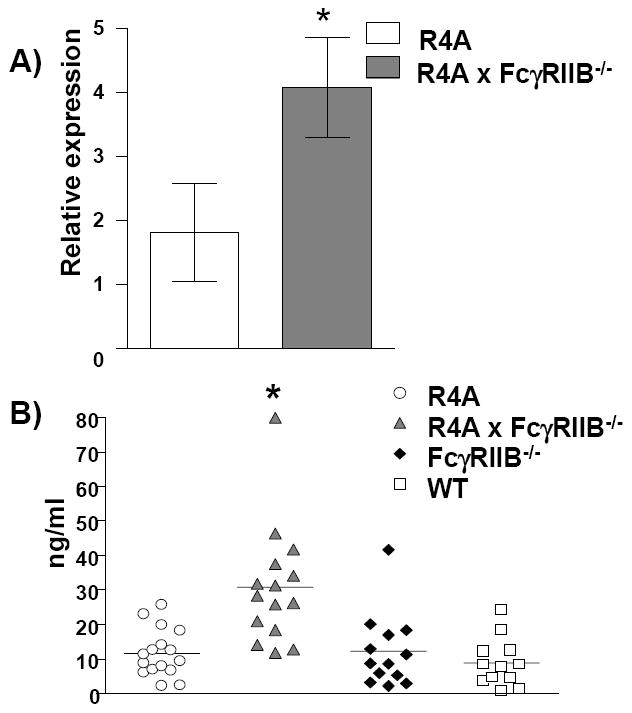
Figure 5. Induction of BAFF.

A) Splenocytes were isolated from 5 to 10 month-old R4A mice or R4A × FcγRIIB-/- mice (n = 6 for each) and analyzed for baff mRNA by qPCR. The data were normalized to polr2a which revealed an increase in baff mRNA in R4A × FcγRIIB-/- mice. B) Serum BAFF levels were measured by ELISA for aged-matched WT BALB/c (n = 12), FcγRIIB-/- (n = 13), R4A (n = 16) and R4A × FcγRIIB-/- (n = 15) mice. **BAFF serum levels were significantly increased in R4A × FcγRIIB-/- mice compared to R4A (p < 0.0001), FcγRIIB-/- (p < 0.0008) or WT (p < 0.0001) BALB/c mice. Statistical significance was determined by Mann-Whitney. Data are shown as the mean ± SD.
