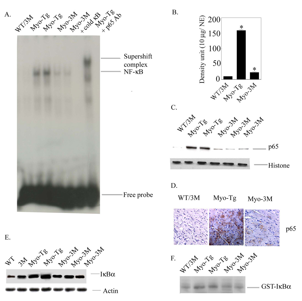
Figure 2. NF-κB activation cascades Myo-3M mice hearts.
(A) Nuclear protein was extracted from the hearts of WT/3M, Myo-Tg mice and Myo-3M. Binding reactions were performed with an NF-κB oligonucleotide labeled with 32P-dATP. The complex formation was eliminated with excess unlabeled NF-κB oligonucleotide. The complex formation was confirmed by supershift analysis using p65 antibody. NE: Nuclear extract. (B) Quantification of EMSA using an arbitrary density unit (10 µg/NE). (C) Western blots profile of NF-κB p65 protein in the nucleus. Histone antibody was used as an internal nuclear protein loading control. (D) Expression of p65 active protein in the heart section of both Myo-Tg and Myo-3M mice and were photographed with an Olympus photomicroscope at 20 × magnification. This figure is representative of three different mice in each group (WT/3M and Myo-Tg). (E). Cytoplasmic protein extracts were made from both WT, 3M, Myo-Tg and Myo-3M mouse hearts at 24 weeks of age. Tissue extracts (50 µg) were analyzed for the intracellular level of total IκBα protein content and (F) Actin protein was used as an internal loading control. Results are presented as the mean SEM and represent three different mice in each group (Myo-Tg and Myo-3M (p < 0.001).

