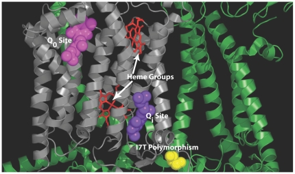
Figure 5. Cytochrome b within the cytochrome bc1 complex.
Only one monomer is shown. The cytochrome b subunit is shown in grey with surrounding subunits shown in green. The CoQ coenzymes are shown bound in the outside (Qo) and inside (Qi) binding sites. The heme groups that are involved in the transfer of the electrons between binding sites are also shown. The cytbI7T polymorphism (yellow) is located on the N-terminus tail of cytb near the Qi site. The three-dimensional coordinates of the complex were obtained from the protein data bank (http://www.pdb.org) [43] under the entry 1ntz [35]. The structure was visually rendered with PyMOL [44].

